Small Bowel Endoscopy Breakthroughs
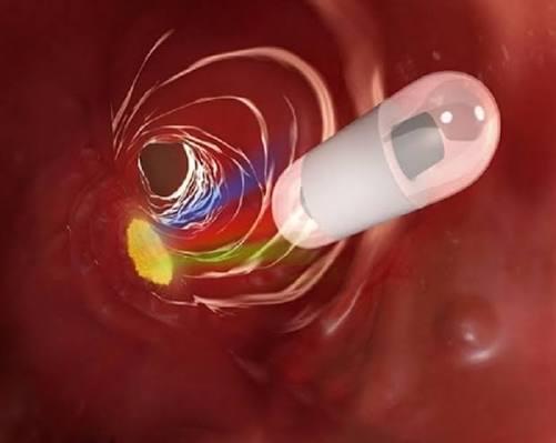
Small bowel endoscopy has long been the "holy grail" of gastroenterology because the small intestine is tortuous, long, and inaccessible to standard upper or lower endoscopes. The advent of video capsule technology solved this problem. Today, Small Bowel Endoscopy primarily refers to capsule endoscopy, though device-assisted enteroscopy exists for therapeutic cases. Patients swallow a vitamin-sized capsule that streams real-time color images, allowing physicians to diagnose celiac disease, obscure bleeding, and small bowel tumors without surgery.
Why Traditional Methods Failed
Before capsule technology, doctors relied on barium follow-throughs and CT enterography, which had low sensitivity for flat lesions. Push enteroscopy could only reach the proximal jejunum. Intraoperative endoscopy was invasive. Wireless Capsule Imaging changed everything by passively traversing the entire 6–8 meters of small bowel. The first human use was reported in 2000, and within five years, capsule endoscopy became the gold standard for obscure GI bleeding.
The Procedure Step by Step
Preparation includes a clear liquid diet and sometimes simethicone. The patient ingests the capsule with water in a clinic. A sensor array attached to the abdomen captures signals from the capsule. Over 8 hours, the patient returns, and the recorder is removed. The physician then views the 50,000–60,000 images at variable speeds. Abnormalities are flagged using software that highlights vascular lesions, ulcerations, or masses. This non-invasive approach has increased patient compliance dramatically.
Clinical Case Example
A 65-year-old with recurrent iron-deficiency anemia had three negative colonoscopies and two gastroscopies. Small bowel endoscopy using a capsule revealed a small angiodysplasia in the mid-ileum. Argon plasma coagulation via double-balloon enteroscopy later treated the lesion. Without capsule imaging, this patient would have continued receiving blood transfusions indefinitely.
Emerging Innovations
Artificial intelligence is now being integrated to automatically detect polyps and bleeding. Additionally, robotic capsules that can stop, move, and take biopsies are in development. These advances will make small bowel endoscopy even more powerful.
In conclusion, Small Bowel Endoscopy combined with wireless capsule imaging provides a complete diagnostic pathway. As capsule technology improves, the need for invasive exploratory surgery will continue to decline. For patients and physicians alike, this represents a win-win: more information, less discomfort, and faster recovery.
Kategorien
Mehr lesen
During a recent presentation at Syscan in Singapore, renowned security researcher and Pwn2Own champion Charlie Miller revealed a serious flaw affecting iPhones. This vulnerability enables attackers to execute malicious code remotely via SMS messages. Such exploits could grant access to sensitive device features like GPS and microphones, potentially revealing the user's location or allowing...
Navigating Netflix's endless catalog can turn choice into a challenge. The paradox of abundance often leaves viewers scrolling rather than watching. This is where the Top 10 list becomes a vital guide, cutting through the clutter. It reveals the collective pulse of the audience, highlighting what's truly capturing attention. Currently, a fresh extraterrestrial suspense film has seized the peak...
Kurz vor Weihnachten ist die Auswahl für das EA FC 26 Team der Woche 15 eher überschaubar, da die meisten Ligen bereits ihre letzten Spiele absolviert haben und in die Winterpause gehen. Einige Ligen, wie die Ligue 1, befinden sich bereits im Stillstand. Dadurch gab es in den letzten Spielrunden nur wenige Partien, was die Kandidaten für das TOTW 15 einschränkt. Trotzdem...
With the Lord of Hatred expansion now live for several weeks, Maxroll’s Diablo IV coverage has been fully refreshed. The Build Guides have been thoroughly updated to incorporate new items, features, and mechanic changes, and the Tier Lists are now complete across all major game modes. Tier List Categories The tier lists are provided in five key areas: Overall Endgame, Bossing, Pit Push,...
Netflix May 2016 Departures: Last Chance to Stream These Titles As May approaches, Netflix subscribers should be aware of approximately 70 movies and TV series that will soon be unavailable on the streaming platform. Here's your final opportunity to catch these departing titles before they vanish! Early May Departures (May 1-7) Several classic films will exit on May 1, including Stanley...